
(April 29, 2021) at 40€")
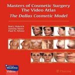
Masters of Cosmetic Surgery The Video Atlas The Dallas Cosmetic Model (Original PDF from Publisher+Videos)(April 29, 2021) at 40€
1,100,000 تومان
اين مجموعه بصورت فايل هاي ويديويي همراه ایبوک است که بر روي دی وی دی يا فلش ارسال ميشود.
مجموعه کامل از سايت اصلي خريداري شده است
پس از سفارش این محصول، از طریق پشتیبانی سایت با شما تماس گرفته میشود و محصول براتون ارسال می گردد.

Masters of Cosmetic Surgery – The Video Atlas: The Dallas Cosmetic Model (Original PDF from Publisher+Videos)(April 29, 2021)
€40.00
Publisher PDF+MP4 (109 High Quality Videos)
Quickly learn and master essential aesthetic surgical procedures from global experts!
The rapid growth in global demand for cosmetic surgery has led to an urgent need for aesthetic surgeons to learn an ever-growing menu of advanced procedures. Masters of Cosmetic Surgery-The Video Atlas: The Dallas Cosmetic Model edited by internationally renowned plastic surgeon Rod J. Rohrich and esteemed colleagues Sammy Sinno and Paul N. Afrooz presents an amazing new method of learning cosmetic surgery techniques. The didactic video guide features contributions from a Who’s Who of superb surgeons and dermatologists that are committed to excellence in their own practices and educational endeavors.
Thirteen sections and 93 succinct chapters are brought to life through key video segments in each chapter that expound on how to perform procedures safely and efficiently to achieve optimal outcomes. This unique resource covers 90 procedures and includes more than 12 hours of video, providing clinicians with the ability to read, see, and hear from an impressive cadre of global experts. Procedural chapters cover the face and neck; nose; eyelids, brow, and forehead; ears; lips; chin and jaw; neuromodulators; fillers; facial resurfacing; breast augmentation and reduction; body contouring; vaginal rejuvenation; and non-surgical body contouring.
Key Highlights
- Masters in cosmetic medicine and surgery share clinical pearls on how to flawlessly perform procedures and optimally handle practice management issues
- Short videos provide an easy method for seeing, performing, and perfecting procedures, resulting in greater efficiency, skill, and safety
- Well-illustrated high-yield text including key points, preoperative considerations, anatomical features, step-by-step operative guides, and algorithms enhance the ability to quickly learn key concepts for any procedure
This how and why guide is transformative in its teaching and learning methods, making it the quintessential reference for trainee and board-certified plastic surgeons, dermatologists, facial plastic surgeons, and oculoplastic surgeons.
Videos
| Video 1.1 | The consult. |
| Video 3.1 | How social media has changed all of plastic surgery. |
| Video 4.1 | Social media; the dos and don’ts. This video demonstrates responsible and successful social media use in plastic surgery as outlined in the chapter. Comprehensive facial analysis. A systematic approach to the facial analysis begins from the vertical and horizontal proportions and progresses to each facial subunit in a top-down approach. Detailed facial analysis will help determine proper treatment plan and help educate patients. Facelift and necklift incisions. Understanding the individual subunits of the facelift incision will help the surgeon to create a well-concealed scar for both male and female patients. Extended SMAS technique. This video demonstrates the key technical components of the extended SMAS technique as outlined in the chapter. SMASectomy. |
| Video 5.1 | |
| Video 6.1 | |
| Video 7.1 | |
| Video 8.1 | |
| Video 9.1 | The lift and fill face lift—autologous fat grafting |
| Video 10.1 | Facelift technique. |
| Video 11.1 | SMAS plication with extended platysma-SMAS flap. |
| Video 12.1 | High SMAS facelift. |
| Video 13.1 | Facial fat grafting. This 57-year-old woman presented improved volume in her face. This video demonstrates the key technical components of this procedure as outlined in the chapter. Clinical nasal analysis. This 63-year-old woman presented for a rhinoplasty consultation. This video demonstrates the key components of the rhinoplasty consultation as outlined in the chapter. Open rhinoplasty approach. |
| Video 14.1 | |
| Video 15.1 | |
| Video 15.2 | Open approach to rhinoplasty. |
| Video 15.3 | Inferior turbinate microfracture. |
| Video 15.4 | Septal access and reconstruction. |
| Video 15.5 | Component dorsal reduction. |
| Video 15.6 | Low-to-low osteotomy. |
| Video 15.7 | Tip shaping endpoints. |
| Video 16.1 | The sequential steps of closed rhinoplasty. This tip rhinoplasty video demonstrates preoperative preparation, incisions, skeletonization, elevation of depressor septi nasi, septal cartilage harvest, tip delivery, cephalic trim, tip suturing, columellar strut placement, closure, alar sill reduction, and splinting. Graduated approach to tip projection. This 49-year-old man presented for a primary rhinoplasty. This video demonstrates the key technical components of nasal tip projection as outlined in the chapter. Bulbous and boxy tip correction. This video reviews the critical aspects for determining how to correct the various morphologies of bulbous and boxy tips during rhinoplasty. Repositioning of the caudal septum. |
| Video 17.1 | |
| Video 18.1 | |
| Video 19.1 | |
| Video 19.2 | Septal rotation suture. |
| Video 20.1 | The ethnic nose. |
| Video 21.1 | Preservation rhinoplasty: Dorsal preservation. |
| Video 22.1 | Spreader grafts. |
| Video 23.1 | Four-step spreader flap technique. |
| Video 24.1 | Columellar strut graft. |
| Video 25.1 | Primary open rhinoplasty. |
| Video 26.1 | Caudal septal extension graft with extended spreaders. |
xxvii
| Video 27.1 | Alar base surgery. Understanding the various morphologies of the alar base will allow the surgeon to ap propriately determine the type of excision required to correct the flare morphologies. This video reviews a systematic approach to marking the patient and performing alar flare reduction. Revision rhinoplasty. This 41-year-old woman presented for a revision rhinoplasty. This video demonstrates the key technical components of this procedure as outlined in the chapter. Periorbital evaluation. This video demonstrates a systematic approach for performing the periorbital evaluation in preparation for upper and/or lower eyelid blepharoplasty. Upper eyelid blepharoplasty. This is a review of the five-step upper eyelid blepharoplasty as described by the senior author. The five-step lower blepharoplasty technique. This 58-year-old woman presented for an improvement in her lower eyelids. This video demonstrates the key technical components of this procedure as outlined in the chapter. Upper eyelid fat grafting. |
| Video 28.1 | |
| Video 29.1 | |
| Video 30.1 | |
| Video 31.1 | |
| Video 32.1 | |
| Video 33.1 | Forehead rejuvenation. |
| Video 34.1 | Endoscopic temporal brow lift. |
| Video 35.1 | Forehead and temple fat augmentation. |
| Video 36.1 | Simplified lateral brow lift. |
| Video 37.1 | Subcutaneous temporal brow lift. |
| Video 38.1 | Otoplasty. |
| Video 39.1 | Surgical correction of prominent ears. This video demonstrates the senior author’s surgical technique for evaluating and correcting prominent ear deformity. Lip lift. |
| Video 40.1 | |
| Video 40.2 | Lip lift. |
| Video 41.1 | Lip fillers. |
| Video 41.2 | Lip injection. |
| Video 42.1 | Technique for fat harvest. Usually, the senior author harvests autologous fat from the inner thigh and then prepares it for injection using centrifugation. Technique for perioral augmentation. In this video, the senior author first volumizes the lateral hollowing followed by augmentation of the upper and lower perioral regions. Perioral neuromodulation. |
| Video 42.2 | |
| Video 43.1 | |
| Video 44.1 | Jawline filler technique. Recorded demonstration of a cannula filler treatment for the filler treatment of a patient’s cheeks, administered by facial plastic surgeon K. Kay Durairaj, MD, FACS. Chin augmentation with permanent silicone implant. |
| Video 45.1 | |
| Video 46.1 | General fat harvest technique. |
| Video 46.2 | Chin augmentation with autologous fat grafting. |
| Video 47.1 | Anatomy dissection and operative examples of buccal fat excision. |
| Video 48.1 | Neuromodulation injection: Forehead and glabella. |
| Video 49.1 | Botulinum toxin injection to crow’s feet. |
| Video 50.1 | Microbotox of the face and neck. |
| Video 51.1 | Neck bands. This 63-year-old woman presented for an improvement in her neck bands. This video demonstrates the key technical components of this procedure as outlined in the chapter. Neurotoxin use for masseter hypertrophy. |
| Video 52.1 | |
| Video 53.1 | Filler finesse: Forehead and temple. |
| Video 54.1 | Temple injection with hyaluronic acid. A 45-year-old woman with hollow temples seen 4 months after injection. Note the apparent saline overfill which absorbs quickly. |
Video 55A.1 Malar augmentation. This 42-year-old woman presented for increased cheek volume. This video
demonstrates the key technical components of this procedure as outlined in the chapter.
Videos
xxviii
Video 55B.1 Filler finesse: Cheek injection technique.
| Video 56.1 | Filler finesse: Tear trough and upper eyelid. |
| Video 57A.1 Filler finesse: Nose. This 39-year-old woman presented for nasal augmentation using filler. This video | |
| demonstrates the key technical components of this procedure as outlined in the chapter. | |
| Video 57B.1 Structural approach to nonsurgical rhinoplasty. | |
| Video 58.1 | Filler finesse: Upper eyelid sulcus. A 48-year-old female 2 weeks after brow injections and 5 days after temple fill. Filler finesse: Hands—Cannula technique with Radiesse. |
| Video 59.1 | |
| Video 59.2 | The five-step hand rejuvenation: Filling with hyaluronic acid. |
| Video 60.1 | TCA peel. |
| Video 61.1 | Dermabrasion of the face. |
| Video 62.1 | Laser resurfacing. Ablative fractional CO2 resurfacing for acne scarring: Consultation, treatment, and post-treatment considerations. Microneedling. Pretreatment and post-treatment set-up with clinical demonstration of microneedling treatment of perioral rhytides, striae distensae, acne, and surgical scars. Breast augmentation. Video of a four-part, dual-plane breast augmentation on a 34-year-old female. Herein we illustrate tissue-based planning, preoperative markings, refine surgical techniques, and postoperative photos. Surgical technique for performing subfascial breast augmentation. |
| Video 63.1 | |
| Video 64.1 | |
| Video 65.1 | |
| Video 66.1 | Surgical technique for performing vertical scar mastopexy with autoaugmentation flap. |
| Video 67.1 | A step-by-step guide to mastopexy via superior pedicle, top down, minus-plus technique with auto-augmentation and fat grafting. The author’s surgical pearls in augmentation mastopexy. |
| Video 68.1 | |
| Video 69.1 | This video shows preoperative markings and surgical maneuvers from start to finish for a typical mastopexy augmentation. The patient presented with ptosis and deflation after breast feeding. The approach was a classic vertical mastopexy with the T added at the end to accommodate the implant |
height after augmentation. In our practice we refer to this pattern as the “owl-shaped” incision, as the
outline appears as an owl with feet. The “feet” is represented by the adjustable horizontal incision to
correct the inferior breast length at the end of the procedure.
| Video 70.1 | Augmentation mastopexy. |
| Video 71.1 | Surgical technique for performing vertical scar breast reduction. |
| Video 72.1 | Wise pattern breast reduction markings and key operative steps. |
| Video 73.1 | Video showing a four-part, dual-plane breast augmentation on a 25-year-old female with constricted lower pole of the breast. This includes tissue-based planning, preoperative markings, refine surgical technique, and postoperative photos. Circumferential SAFE lipoplasty with gluteal augmentation. |
| Video 74.1 | |
| Video 75.1 | This 36-year-old man presented for high-definition abdominal liposculpture with BodyBanking. This video demonstrates the key technical components of this procedure as outlined in the chapter. High-definition lipoplasty. |
| Video 76.1 | |
| Video 77.1 | Male high-definition liposculpture. |
| Video 78.1 | This 27-year-old man presented for gynecomastia excision with BodyBanking. He complained of gynecomastia. This video demonstrates the key technical components of gynecomastia excision with BodyBanking as outlined in the chapter. Mommy makeover finesse. |
| Video 79.1 | |
| Video 80.1 | Brachioplasty refinements. |
| Video 81.1 | Medial thigh lift. |
| Video 82.1 | Postbariatric brachioplasty markings and key operative steps. |
Videos
xxix
| Video 83.1 | Lower body lift. The patient is a 46-year-old female with a history of 100-lb weight loss with lifestyle modification. Preoperative BMI was 26.7 kg/m2. She desired a lower body lift with autoaugmentation. Step-by-step execution of vertical thigh lift using the avulsion thighplasty technique. |
| Video 84.1 | |
| Video 85.1 | Bra-line back lift. |
| Video 86.1 | Buttock augmentation: S-curve. |
| Video 87.1 | This 32-year-old woman presented for Brazilian butt lift (BBL). She complained of inadequate projection and excess fat in the waist, abdomen, and back. This video demonstrates the key technical components of SSBA as outlined in the chapter. Labiaplasty with Aviva. |
| Video 88.1 | |
| Video 89.1 | Bipolar radiofrequency treatment of labia majora and minora. |
| Video 89.2 | Fractional radiofrequency treatment of mons and labia majora. |
| Video 90.1 | Cryolipolysis. |
| Video 91.1 | Marking for lower face and neck bipolar radiofrequency. |
| Video 91.2 | Fractional radiofrequency using Morpheus8. |
| Video 91.3 | An intraoperative example of fractional radiofrequency. |
| Video 92.1 | Minimally invasive upper arm contouring with bipolar radiofrequency assisted liposuction. |
| Video 93.1 | Demonstration of the submental markings used to identify the six regions of the expanded safe zone (ESZ). |
How to Buy
Thanks For Choosing WWW.BAHARMEDBOOK.COM .
for buying materials from WWW.BAHARMEDBOOK.COM , Follow the instructions :
Step 1 : Contact us :
Email : INFO@BAHARMEDBOOK.COM
Telegram : @BAHARMEDBOOKADMIN
INSTAGRAM : https://www.instagram.com/bahar.medbooks
WhatsApp : +98 9123309803
Step 2 : Send Material You Want to Buy
Step 3 : You Will Receieve Payment Invoice
Step 4 : After Payment confirmation , You will Receive Download Link .
Prices: contact us | info@baharmedbook.com
IF your course was not available in our archive, please request it via request form
- توضیحات
- توضیحات
- نظرات (0)
توضیحات
اين مجموعه بصورت فايل هاي ويديويي همراه ایبوک است که بر روي دی وی دی يا فلش ارسال ميشود.
مجموعه کامل از سايت اصلي خريداري شده است
پس از سفارش این محصول، از طریق پشتیبانی سایت با شما تماس گرفته میشود و محصول براتون ارسال می گردد.
Masters of Cosmetic Surgery – The Video Atlas: The Dallas Cosmetic Model (Original PDF from Publisher+Videos)(April 29, 2021)
€40.00
Publisher PDF+MP4 (109 High Quality Videos)
Quickly learn and master essential aesthetic surgical procedures from global experts!
The rapid growth in global demand for cosmetic surgery has led to an urgent need for aesthetic surgeons to learn an ever-growing menu of advanced procedures. Masters of Cosmetic Surgery-The Video Atlas: The Dallas Cosmetic Model edited by internationally renowned plastic surgeon Rod J. Rohrich and esteemed colleagues Sammy Sinno and Paul N. Afrooz presents an amazing new method of learning cosmetic surgery techniques. The didactic video guide features contributions from a Who’s Who of superb surgeons and dermatologists that are committed to excellence in their own practices and educational endeavors.
Thirteen sections and 93 succinct chapters are brought to life through key video segments in each chapter that expound on how to perform procedures safely and efficiently to achieve optimal outcomes. This unique resource covers 90 procedures and includes more than 12 hours of video, providing clinicians with the ability to read, see, and hear from an impressive cadre of global experts. Procedural chapters cover the face and neck; nose; eyelids, brow, and forehead; ears; lips; chin and jaw; neuromodulators; fillers; facial resurfacing; breast augmentation and reduction; body contouring; vaginal rejuvenation; and non-surgical body contouring.
Key Highlights
- Masters in cosmetic medicine and surgery share clinical pearls on how to flawlessly perform procedures and optimally handle practice management issues
- Short videos provide an easy method for seeing, performing, and perfecting procedures, resulting in greater efficiency, skill, and safety
- Well-illustrated high-yield text including key points, preoperative considerations, anatomical features, step-by-step operative guides, and algorithms enhance the ability to quickly learn key concepts for any procedure
This how and why guide is transformative in its teaching and learning methods, making it the quintessential reference for trainee and board-certified plastic surgeons, dermatologists, facial plastic surgeons, and oculoplastic surgeons.
Videos
| Video 1.1 | The consult. |
| Video 3.1 | How social media has changed all of plastic surgery. |
| Video 4.1 | Social media; the dos and don’ts. This video demonstrates responsible and successful social media use in plastic surgery as outlined in the chapter. Comprehensive facial analysis. A systematic approach to the facial analysis begins from the vertical and horizontal proportions and progresses to each facial subunit in a top-down approach. Detailed facial analysis will help determine proper treatment plan and help educate patients. Facelift and necklift incisions. Understanding the individual subunits of the facelift incision will help the surgeon to create a well-concealed scar for both male and female patients. Extended SMAS technique. This video demonstrates the key technical components of the extended SMAS technique as outlined in the chapter. SMASectomy. |
| Video 5.1 | |
| Video 6.1 | |
| Video 7.1 | |
| Video 8.1 | |
| Video 9.1 | The lift and fill face lift—autologous fat grafting |
| Video 10.1 | Facelift technique. |
| Video 11.1 | SMAS plication with extended platysma-SMAS flap. |
| Video 12.1 | High SMAS facelift. |
| Video 13.1 | Facial fat grafting. This 57-year-old woman presented improved volume in her face. This video demonstrates the key technical components of this procedure as outlined in the chapter. Clinical nasal analysis. This 63-year-old woman presented for a rhinoplasty consultation. This video demonstrates the key components of the rhinoplasty consultation as outlined in the chapter. Open rhinoplasty approach. |
| Video 14.1 | |
| Video 15.1 | |
| Video 15.2 | Open approach to rhinoplasty. |
| Video 15.3 | Inferior turbinate microfracture. |
| Video 15.4 | Septal access and reconstruction. |
| Video 15.5 | Component dorsal reduction. |
| Video 15.6 | Low-to-low osteotomy. |
| Video 15.7 | Tip shaping endpoints. |
| Video 16.1 | The sequential steps of closed rhinoplasty. This tip rhinoplasty video demonstrates preoperative preparation, incisions, skeletonization, elevation of depressor septi nasi, septal cartilage harvest, tip delivery, cephalic trim, tip suturing, columellar strut placement, closure, alar sill reduction, and splinting. Graduated approach to tip projection. This 49-year-old man presented for a primary rhinoplasty. This video demonstrates the key technical components of nasal tip projection as outlined in the chapter. Bulbous and boxy tip correction. This video reviews the critical aspects for determining how to correct the various morphologies of bulbous and boxy tips during rhinoplasty. Repositioning of the caudal septum. |
| Video 17.1 | |
| Video 18.1 | |
| Video 19.1 | |
| Video 19.2 | Septal rotation suture. |
| Video 20.1 | The ethnic nose. |
| Video 21.1 | Preservation rhinoplasty: Dorsal preservation. |
| Video 22.1 | Spreader grafts. |
| Video 23.1 | Four-step spreader flap technique. |
| Video 24.1 | Columellar strut graft. |
| Video 25.1 | Primary open rhinoplasty. |
| Video 26.1 | Caudal septal extension graft with extended spreaders. |
xxvii
| Video 27.1 | Alar base surgery. Understanding the various morphologies of the alar base will allow the surgeon to ap propriately determine the type of excision required to correct the flare morphologies. This video reviews a systematic approach to marking the patient and performing alar flare reduction. Revision rhinoplasty. This 41-year-old woman presented for a revision rhinoplasty. This video demonstrates the key technical components of this procedure as outlined in the chapter. Periorbital evaluation. This video demonstrates a systematic approach for performing the periorbital evaluation in preparation for upper and/or lower eyelid blepharoplasty. Upper eyelid blepharoplasty. This is a review of the five-step upper eyelid blepharoplasty as described by the senior author. The five-step lower blepharoplasty technique. This 58-year-old woman presented for an improvement in her lower eyelids. This video demonstrates the key technical components of this procedure as outlined in the chapter. Upper eyelid fat grafting. |
| Video 28.1 | |
| Video 29.1 | |
| Video 30.1 | |
| Video 31.1 | |
| Video 32.1 | |
| Video 33.1 | Forehead rejuvenation. |
| Video 34.1 | Endoscopic temporal brow lift. |
| Video 35.1 | Forehead and temple fat augmentation. |
| Video 36.1 | Simplified lateral brow lift. |
| Video 37.1 | Subcutaneous temporal brow lift. |
| Video 38.1 | Otoplasty. |
| Video 39.1 | Surgical correction of prominent ears. This video demonstrates the senior author’s surgical technique for evaluating and correcting prominent ear deformity. Lip lift. |
| Video 40.1 | |
| Video 40.2 | Lip lift. |
| Video 41.1 | Lip fillers. |
| Video 41.2 | Lip injection. |
| Video 42.1 | Technique for fat harvest. Usually, the senior author harvests autologous fat from the inner thigh and then prepares it for injection using centrifugation. Technique for perioral augmentation. In this video, the senior author first volumizes the lateral hollowing followed by augmentation of the upper and lower perioral regions. Perioral neuromodulation. |
| Video 42.2 | |
| Video 43.1 | |
| Video 44.1 | Jawline filler technique. Recorded demonstration of a cannula filler treatment for the filler treatment of a patient’s cheeks, administered by facial plastic surgeon K. Kay Durairaj, MD, FACS. Chin augmentation with permanent silicone implant. |
| Video 45.1 | |
| Video 46.1 | General fat harvest technique. |
| Video 46.2 | Chin augmentation with autologous fat grafting. |
| Video 47.1 | Anatomy dissection and operative examples of buccal fat excision. |
| Video 48.1 | Neuromodulation injection: Forehead and glabella. |
| Video 49.1 | Botulinum toxin injection to crow’s feet. |
| Video 50.1 | Microbotox of the face and neck. |
| Video 51.1 | Neck bands. This 63-year-old woman presented for an improvement in her neck bands. This video demonstrates the key technical components of this procedure as outlined in the chapter. Neurotoxin use for masseter hypertrophy. |
| Video 52.1 | |
| Video 53.1 | Filler finesse: Forehead and temple. |
| Video 54.1 | Temple injection with hyaluronic acid. A 45-year-old woman with hollow temples seen 4 months after injection. Note the apparent saline overfill which absorbs quickly. |
Video 55A.1 Malar augmentation. This 42-year-old woman presented for increased cheek volume. This video
demonstrates the key technical components of this procedure as outlined in the chapter.
Videos
xxviii
Video 55B.1 Filler finesse: Cheek injection technique.
| Video 56.1 | Filler finesse: Tear trough and upper eyelid. |
| Video 57A.1 Filler finesse: Nose. This 39-year-old woman presented for nasal augmentation using filler. This video | |
| demonstrates the key technical components of this procedure as outlined in the chapter. | |
| Video 57B.1 Structural approach to nonsurgical rhinoplasty. | |
| Video 58.1 | Filler finesse: Upper eyelid sulcus. A 48-year-old female 2 weeks after brow injections and 5 days after temple fill. Filler finesse: Hands—Cannula technique with Radiesse. |
| Video 59.1 | |
| Video 59.2 | The five-step hand rejuvenation: Filling with hyaluronic acid. |
| Video 60.1 | TCA peel. |
| Video 61.1 | Dermabrasion of the face. |
| Video 62.1 | Laser resurfacing. Ablative fractional CO2 resurfacing for acne scarring: Consultation, treatment, and post-treatment considerations. Microneedling. Pretreatment and post-treatment set-up with clinical demonstration of microneedling treatment of perioral rhytides, striae distensae, acne, and surgical scars. Breast augmentation. Video of a four-part, dual-plane breast augmentation on a 34-year-old female. Herein we illustrate tissue-based planning, preoperative markings, refine surgical techniques, and postoperative photos. Surgical technique for performing subfascial breast augmentation. |
| Video 63.1 | |
| Video 64.1 | |
| Video 65.1 | |
| Video 66.1 | Surgical technique for performing vertical scar mastopexy with autoaugmentation flap. |
| Video 67.1 | A step-by-step guide to mastopexy via superior pedicle, top down, minus-plus technique with auto-augmentation and fat grafting. The author’s surgical pearls in augmentation mastopexy. |
| Video 68.1 | |
| Video 69.1 | This video shows preoperative markings and surgical maneuvers from start to finish for a typical mastopexy augmentation. The patient presented with ptosis and deflation after breast feeding. The approach was a classic vertical mastopexy with the T added at the end to accommodate the implant |
height after augmentation. In our practice we refer to this pattern as the “owl-shaped” incision, as the
outline appears as an owl with feet. The “feet” is represented by the adjustable horizontal incision to
correct the inferior breast length at the end of the procedure.
| Video 70.1 | Augmentation mastopexy. |
| Video 71.1 | Surgical technique for performing vertical scar breast reduction. |
| Video 72.1 | Wise pattern breast reduction markings and key operative steps. |
| Video 73.1 | Video showing a four-part, dual-plane breast augmentation on a 25-year-old female with constricted lower pole of the breast. This includes tissue-based planning, preoperative markings, refine surgical technique, and postoperative photos. Circumferential SAFE lipoplasty with gluteal augmentation. |
| Video 74.1 | |
| Video 75.1 | This 36-year-old man presented for high-definition abdominal liposculpture with BodyBanking. This video demonstrates the key technical components of this procedure as outlined in the chapter. High-definition lipoplasty. |
| Video 76.1 | |
| Video 77.1 | Male high-definition liposculpture. |
| Video 78.1 | This 27-year-old man presented for gynecomastia excision with BodyBanking. He complained of gynecomastia. This video demonstrates the key technical components of gynecomastia excision with BodyBanking as outlined in the chapter. Mommy makeover finesse. |
| Video 79.1 | |
| Video 80.1 | Brachioplasty refinements. |
| Video 81.1 | Medial thigh lift. |
| Video 82.1 | Postbariatric brachioplasty markings and key operative steps. |
Videos
xxix
| Video 83.1 | Lower body lift. The patient is a 46-year-old female with a history of 100-lb weight loss with lifestyle modification. Preoperative BMI was 26.7 kg/m2. She desired a lower body lift with autoaugmentation. Step-by-step execution of vertical thigh lift using the avulsion thighplasty technique. |
| Video 84.1 | |
| Video 85.1 | Bra-line back lift. |
| Video 86.1 | Buttock augmentation: S-curve. |
| Video 87.1 | This 32-year-old woman presented for Brazilian butt lift (BBL). She complained of inadequate projection and excess fat in the waist, abdomen, and back. This video demonstrates the key technical components of SSBA as outlined in the chapter. Labiaplasty with Aviva. |
| Video 88.1 | |
| Video 89.1 | Bipolar radiofrequency treatment of labia majora and minora. |
| Video 89.2 | Fractional radiofrequency treatment of mons and labia majora. |
| Video 90.1 | Cryolipolysis. |
| Video 91.1 | Marking for lower face and neck bipolar radiofrequency. |
| Video 91.2 | Fractional radiofrequency using Morpheus8. |
| Video 91.3 | An intraoperative example of fractional radiofrequency. |
| Video 92.1 | Minimally invasive upper arm contouring with bipolar radiofrequency assisted liposuction. |
| Video 93.1 | Demonstration of the submental markings used to identify the six regions of the expanded safe zone (ESZ). |
How to Buy
Thanks For Choosing WWW.BAHARMEDBOOK.COM .
for buying materials from WWW.BAHARMEDBOOK.COM , Follow the instructions :
Step 1 : Contact us :
Email : INFO@BAHARMEDBOOK.COM
Telegram : @BAHARMEDBOOKADMIN
INSTAGRAM : https://www.instagram.com/bahar.medbooks
WhatsApp : +98 9123309803
Step 2 : Send Material You Want to Buy
Step 3 : You Will Receieve Payment Invoice
Step 4 : After Payment confirmation , You will Receive Download Link .
Prices: contact us | info@baharmedbook.com
IF your course was not available in our archive, please request it via request form


نقد و بررسیها
هنوز بررسیای ثبت نشده است.