

Musculoskeletal Imaging 2ed PDF+Video at 2€
60,000 تومان
- توضیحات
- توضیحات
- نظرات (0)
توضیحات
Product Details / مشخصات محصول
———————————————————————————-
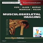
Musculoskeletal Imaging 2ed PDF+Video at 2€
این فایل پی دی اف و فیلم، کامل از سايت اصلي خريداري شده است
به این صورت که
E-chapter
هم در فایل
True PDF
موجود است
و فایل همراه تمام قسمتهای الکترونیکی و فهرست و ایندکس است که فقط در فایل اصلی انتشارات موجود است.
———————————————————————————-
فایل محصولات در سایت موجود نیست و پس از خرید شما در ساعات کاری آپلود و به آدرس و یا ایمیل ارسال و در اختیار شما قرار خواهد گرفت. در صورت بروز مشکل در ساعات اداری پاسخگوی شما خواهیم بود
فايل کتاب فوق در فرمت PDF ، با کيفيت عالي و قابل اجرا و انتقال بر روي کامپيوتر، تبلت و موبايل مي باشد. امکان جستجو و کپي متن و همچنين ذخيره تصاوير از ديگر ويژگي هاي اين محصول مي باشد.
همچنين جهت خريد نسخه چاپي کتاب با شماره 09123309803 تماس بگيريد و يا با کليک بر روي دکمه لینک سفارش ، با ثبت اطلاعات خود منتظر تماس کارشناسان فروش ما باشيد.
وقتی کاربری اقدام به خرید کتاب از سایت می نماید، پس از یافتن کتاب مورد نظر در سایت، می تواند کتاب موردنظرش را با تمام مشخصه های دلخواه خود خرید کند.
یعنی می تواند کتاب را با تعداد جلد دلخواه، در سایز و قطع دلخواه و با نوع صحافی دلخواه خرید کند.
کتاب از نظر نوع صحافی به انواع زیر تقسیم می شود
- جلد سخت یا گالینگور (Hard Cover)
- جلد نرم (Soft Cover)
- سیمی فنری
———————————————————————————-
How to Buy …
you should pay 2.00€ Euro via below payment Link
https://yekpay.me/en/baharmedbook.com
Pay attention to Send Money To abbas kouhiesfahani “baharmedbook manager Bank account”
After Payment confirmation , contact us | info@baharmedbook.com and You will Receive Direct Download Link .
———————————————————————————-
IF your Material was not available in our archive, please request it via request form on www.baharmedbook.com
———————————————————————————-
دانلود کتاب تصویربرداری اسکلتی عضلانی + ویدئو
Musculoskeletal Imaging, 2ed + Video
In its fully revised and updated second edition, Musculoskeletal Imaging covers every aspect of musculoskeletal radiology. This medical reference book incorporates the latest diagnostic modalities and interventional techniques, as well as must-read topics such as hip, groin and cartilage imaging; newly described impingements; and new concepts in the hip including teres ligament pathology. Accessibility in print, online and across portable devices makesMusculoskeletal Imaging a fully searchable and dependable sourcefor both reading and reference. This publication is a key title in the popular Expert Radiology Series, which delivers evidence-based expert guidance from around the globe.
“This is an excellent benchbook and accompanying electronic resource which will be of value to trainee radiologists and established consultants.”Reviewed by: Dr Steve Amerasekara, Consultant Radiologist on behalf of journal RAD Magazine Date: July 2015
“This outstanding text is now an acclaimed primary resource and therefore belongs in the libraries and at the work stations of all general and orthopedic hospital departments of radiology and, indeed, at any and all imaging facilities involved in musculoskeletal imaging.” Foreword by: Lee F. Rogers, June 2015
- Fully understand each topic with a format that delivers essential background information.
- Streamline the decision-making process with integrated protocols, classic signs, and ACR guidelines, as well as a design that structures every chapter consistently to include pathophysiology, imaging techniques, imaging findings, differential diagnosis, and treatment options.
- Write the most comprehensive reports possible with help from boxes highlighting what the referring physician needs to know, as well as suggestions for treatment and future imaging studies.
- Access in-depth case studies, valuable appendices, and additional chapters covering all of the most important musculoskeletal procedures performed today.
- Quickly locate important information with a full-color design that includes color-coded tables and bulleted lists highlighting key concepts, as well as color artwork that lets you easily find critical anatomic views of diseases and injuries.
- Engage with more than 40 brand-new videos, including arthroscopic videos.
- Easily comprehend complicated material with over 5,000 images and new animations.
- Explore integrated clinical perspectives on the newest modalities such as PET-CT in cancer, diffusion MR, as well as ultrasonography, fusion imaging, multi-slice CT and nuclear medicine.
- Learn from team of international experts provides a variety of evidence-based guidance, including the pros and cons of each modality, to help you overcome difficult challenges.
- Expert Consult eBook version included with purchase. This enhanced eBook experience allows you to search all of the text, figures, references, and videos from the book on a variety of devices.
Review
“This is an impressive, comprehensive update of musculoskeletal radiology. It has a disciplined, systematic organization allowing for discussion of a full range of topics, with balanced attention to radiography, ultrasound, MRI, CT, and NM findings. There is a strong focus on clinical application of imaging findings and strong attention to anatomical and pathological detail. Both contemporary and classic findings are included. The ebook format allows for easy reference when in clinical practice. This could serve as the primary resource for a musculoskeletal fellowship.”
–B. Keegan Markhardt, MD (University of Wisconsin-Madison Hospital and Clinics) Doody Score: 97 – ۵ Stars
“This book provides the best of both worlds. There is a 1,291 page paper version. In addition, access to an electronic version (which can be accessed on or offline) is free with paper purchases, or can be bought as a standalone item. I used the online version and an offline version adapted for mobile devices; both worked well and were reasonably intuitive…This is an excellent benchbook and accompanying electronic resource which will be of value to trainee radiologists and established consultants.”
Reviewed by: Dr Steve Amerasekara, Consultant Radiologist on behalf of journal RAD Magazine Date: July 2015
“Musculoskeletal Imaging is an outstanding, definitive work. The authors and publisher have come together to produce a work that maximizes the advantages of both print and electronic media. The editors?Drs. Pope, Bloem, Beltran, Morrison, and Wilson?are to be congratulated and commended for their efforts in producing such a great contribution to the field of radiology.
This outstanding text is now an acclaimed primary resource and therefore belongs in the libraries and at the work stations of all general and orthopedic hospital departments of radiology and, indeed, at any and all imaging facilities involved in musculoskeletal imaging.” Reviewed by: Lee F. Rogers, Feb 2015
Contents
۱. General Imaging Principles
۲. General Principles of Osseous Injury
۳. Imaging of Facial and Skull Trauma
۴. Cervical Spine Injuries
۵. Injury of the Thoracic Cage and Thoracolumbar Spine
۶. Normal Shoulder
۷. Osseous Injuries of the Shoulder Girdle
۸. Shoulder Impingement Syndromes
۹. Glenohumeral Instability
۱۰. Normal Elbow
۱۱. Acute Osseous Injury of the Elbow and Forearm
۱۲. Soft Tissue Injury to the Elbow
۱۳. Normal Wrist
۱۴. Acute Osseous Injury to the Wrist
۱۵. Internal Derangement of the Wrist
More…
۱۱۰. Postoperative Knee
۱۱۱. Postoperative Ankle and Foot
۱۱۲. Imaging of the Residual Limb after Amputation
۱۱۳. Postoperative Infections
۱۱۴. Temporomandibular Joint
۱۱۵. Dental Imaging
۱۱۶. Normal Variants
۱۱۷. Biopsy: Soft Tissue
۱۱۸. Percutaneous Biopsy of the Appendicular Skeleton
۱۱۹. Percutaneous Biopsy of the Spine
۱۲۰. Tumor Ablation
۱۲۱. Spinal Injections
۱۲۲. Discography
۱۲۳. Vertebroplasty and Kyphoplasty
۱۲۴. Percutaneous Intradiskal Therapies
۱۲۵. Ultrasound Procedures
Video Contents
۳-۱. Illustration of Various Types of Facial and Skull Fractures and Mechanisms of Injury.
۳-۲. Fractures of the Orbital Floor with Orbital Emphysema.
۳-۳. Complex Maxillofacial fracture.
۳-۴. “Pyramidal” Fracture of the Orbital Floor and Maxillary Sinuses (Sagittal).
۳-۵. “Pyramidal” Fracture of the Orbital Floor and Maxillary Sinuses (Coronal).
۳-۶. Frontal Sinus Fractures: Axial Bone Window Imaging.
۳-۷. Frontal Sinus Fractures: Axial Soft Tissue Window Imaging.
۳-۸. Bilateral Nasal Bone Fractures.
۳-۹. Depressed Fracture of the Left Orbit Floor.
۳-۱۰. Fractures of the Right Orbit Floor and Lamina Papyracea.
۳-۱۱. Bilateral Comminuted Nasal Bone Fractures.
۳-۱۲. ZMC Fracture Before Repair with Placement of Osteosynthesis Plates.
۳-۱۳. ZMC Fracture Before Repair with Placement of Osteosynthesis Plates (3D Reconstruction).
۳-۱۴. ZMC Fracture After Repair with Placement of Osteosynthesis Plates.
۳-۱۵. ZMC Fracture After Repair with Placement of Osteosynthesis Plates (3D reconstruction).
۳-۱۶. Fractures of the Maxillary Sinus Wall (Axial).
۳-۱۷. Fractures of the Maxillary Sinus Wall (Coronal).
۶-۱. Complete MR Arthrogram Series.
۶-۲. Long Biceps Tendon in Position During Internal and External Rotation.
۶-۳. Normal Arthroscopy of the Shoulder.
۶-۴. Te Six Phases in the Overhead Trowing Motion.
۸-۱. Posterosuperior Shoulder Impingement Syndrome.
۸-۲. Full thickness tear of the supraspinatus.
۸-۳. Partial articular surface tear of the supraspinatus.
۹-۱. Anterior Shoulder Instability.
۱۰-۱. ۳D CT Rendering of the Elbow Joint.
۱۰-۲. Ulnar Nerve Subluxation on Ultrasonography.
۱۲-۱. Ulnar Collateral Ligament Complex Injury in the Trowing Athlete.
۱۲-۲. Complete Distal Biceps Tendon Tear.
۱۲-۳. Snapping Triceps.
۱۲-۴. Common Extensor Tenotomy.
۱۸-۱. Skier’s Tumb.
۲۰-۱. Pelvic Fracture.
۲۱-۱. Athletic Pubalgia.
۲۳-۱. Femoracetabular Impingement (FAI).
۲۴-۱. Normal Anatomy of the Knee: Coronal MR.
۲۴-۲. Normal Anatomy of the Knee: Axial MR.
۲۴-۳. Normal Anatomy of the Knee: Sagittal MR.
۲۷-۱. ACL Tear: Mechanism of Injury.
۲۸-۱. Transient Lateral Patellar Dislocation.
۳۲-۱. Inversion Ankle Injuries.
۳۳-۱. Achilles Tendinosis Sonogram (Short-axis).
۳۳-۲. Achilles Tendinosis Sonogram (Long-axis).
۳۳-۳. Complete Tear of the Achilles Tendon Sonogram.
۳۳-۴. Subluxing Tibialis Posterior Tendon Sonogram.
۳۳-۵. Subluxing Peroneus Longus Tendon Sonogram.
۳۳-۶. Short-axis Ultrasound Movie of Tibialis Posterior Tendinopathy with Color Doppler.
۳۳-۷. Cystic Degeneration in a Dysfunctional Tibialis Posterior Tendon Sonogram.
۳۳-۸. Short-axis Ultrasound Movie of Tibialis Posterior Tendinopathy.
۳۳-۹. Extensor Digitorum Longus Tenosynovitis Sagittal Fast Spin Echo Inversion Recovery with Fat Saturation (A) and Axial Fast Spin Echo Proton Density with Saturation (B) MR Movies.
۳۳-۱۰. Tenosynovitis of the Extensor Digitorum Longus after Ultrasound-Guided Aspiration with Subsequent Injection of Steroid.
۳۳-۱۱. Type 2 Accessory Navicula MR Movie.
۳۳-۱۲. Tibialis Posterior Tendinosis MR Movie.
۳۳-۱۳. Enlarged Peroneal Tubercle on MR.
۳۳-۱۴. Impingement Syndromes of the Ankle.
۳۴-۱. Anterolateral Impingement (Syndesmotic Impingement).
۳۴-۲. Anterolateral Impingement.
۳۴-۳. Anterior Impingement.
۳۴-۴. Anteromedial Impingement.
۳۴-۵. Posterior Ankle Impingement.
۳۴-۶. Unstable Osteochondral Lesion of the Medial Talar Dome (Grifth Grade 4b, Berndt and Harty stage III).
۳۴-۷. Osteochondral Lesion Treatment.
۳۴-۸. Osteochondral Lesion Treatment: Microfracture Technique.
۳۸-۱. Little Leaguer’s Shoulder.
۴۷-۱. Cartilage.
۱۱۴-۱. Temporomandibular Joint Anatomy and Mechanism of Motion.
ناشر : Saunders; 2 edition
سال انتشار : 2015
زبان کتاب : انگلیسی
نوع فایل : MP4 + PDF (کیفیت اصلی)
تعداد صفحات : 2884
(ISBN) شابک : 1455708135
قیمت کتاب درآمازون : $209.88
حجم فایل : 600 MB
———————————————————————————-
Thanks For Choosing WWW.BAHARMEDBOOK.COM .
for buying materials from WWW.BAHARMEDBOOK.COM , Follow the instructions :
Step 1 : Contact us :
Email : INFO@BAHARMEDBOOK.COM
Telegram : @BAHARMEDBOOKADMIN
INSTAGRAM : https://www.instagram.com/bahar.medbooks
WhatsApp : +98 9123309803
Step 2 : Send Material You Want to Buy
Step 3 : You Will Receieve Payment Invoice
Step 4 : After Payment confirmation , You will Receive Download Link .
———————————————————————————-
contact us | info@baharmedbook.com
High Quality Videos
Fast Delivery After Payment
Full Customer Support 24/7
AMBOSS, baharmedbook, Buy Medical Course-Videos, Buy Medical E-Books, Buy Medical Exam Source, Buy Medical Journal, Canada QBank for the MCCQE Part 1 at 15€, CME Videos, Family Medicine, FIRST AID for the USMLE STEP 1 2021, lecturio 2021, Lecturio USMLE Step 2 (2020) at 30€, MCCQE, MCCQE Part 1, medical board video, OET Occupational English Test 2020 Pack, osmosis 2021, QBank for the MCCQE Part 1, Question Bank for the Medical Council of Canada Qualifying Exam Part 1, USMLE, usmle step 1 books, آزمون هاي پزشکي خارج از کشور, امتحانات کانادا و استراليا و امريکا, انتشارات پزشکي, انتشارات کتب پزشکي, تهیه OET Occupational English Test 2020 Pack, خريد کتاب پزشکي با تخفيف, خرید OET Occupational English Test 2020 Pack, خرید بانک سوالات MCCQE Part 1, دانلود کتاب پزشکي, فیلم پزشکی, کتاب پزشکی بهار, کتاب رفرنس پزشکي, مديکال بوک, منابع علوم پزشکیTاشتراک سالانه ژورنال های پزشکی, اشتراک مجلات معتبر پزشکی, خرید اشتراک مجلات پزشکی, ژورنال معتبر پزشکی, مجلات پزشکی


نقد و بررسیها
هنوز بررسیای ثبت نشده است.